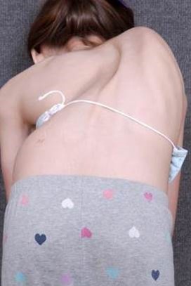
How we correct spine malrotation and rib prominence during spine fusions
3-dimensional problem, with the spine rotating around itself as it bends to the
side. The way to visualize this is to
imagine a water slide, as it turns to the side, it also rotates…just like the
spine in scoliosis and how the spine twists the ribs around.

This post will show how we can improve the spine and rib position during a spine fusion surgery.
The case example is a 14 year old young lady with a 58 degree right thoracic idiopathic curve, and 35 degree proximal thoracic and 24 degree lumbar curves.
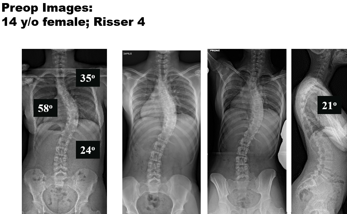
The supine side bending radiographs demonstrate the flexibility of the spine. So only the main thoracic curve of 58 degrees is what we call “structural” and the other two curves, due to the fact they bend out to be below 25 degrees, we call “nonstructural” or “compensatory”.

In order to maximize 3-dimensional correction and long-term outcome, while minimizing need for any additional surgeries the plan was to do a T3 to L1 posterior spinal fusion. T7-T8 and T8-T9 PCOs, or posterior column osteotomies were also performed. These osteotomies (PCOs) are done at the time of surgery to increase spine flexibility in all 3 planes, to maximize spinal deformity correction to as close to “normal” as we can safely achieve.

Pedicle screw are the optimal method of spine fixation. They allow rigid fixation to the spine, and permits the spine to be moved 3-dimensionally. Not every vertebra needs to have 2 screws. Strategically placing screws to optimize immediate correction and assure long-term outcome is preferred, so about 1.5 screws per level is common.
By placing the screws at certain locations, specifically at the apex, the spine can be derotated back more toward normal.
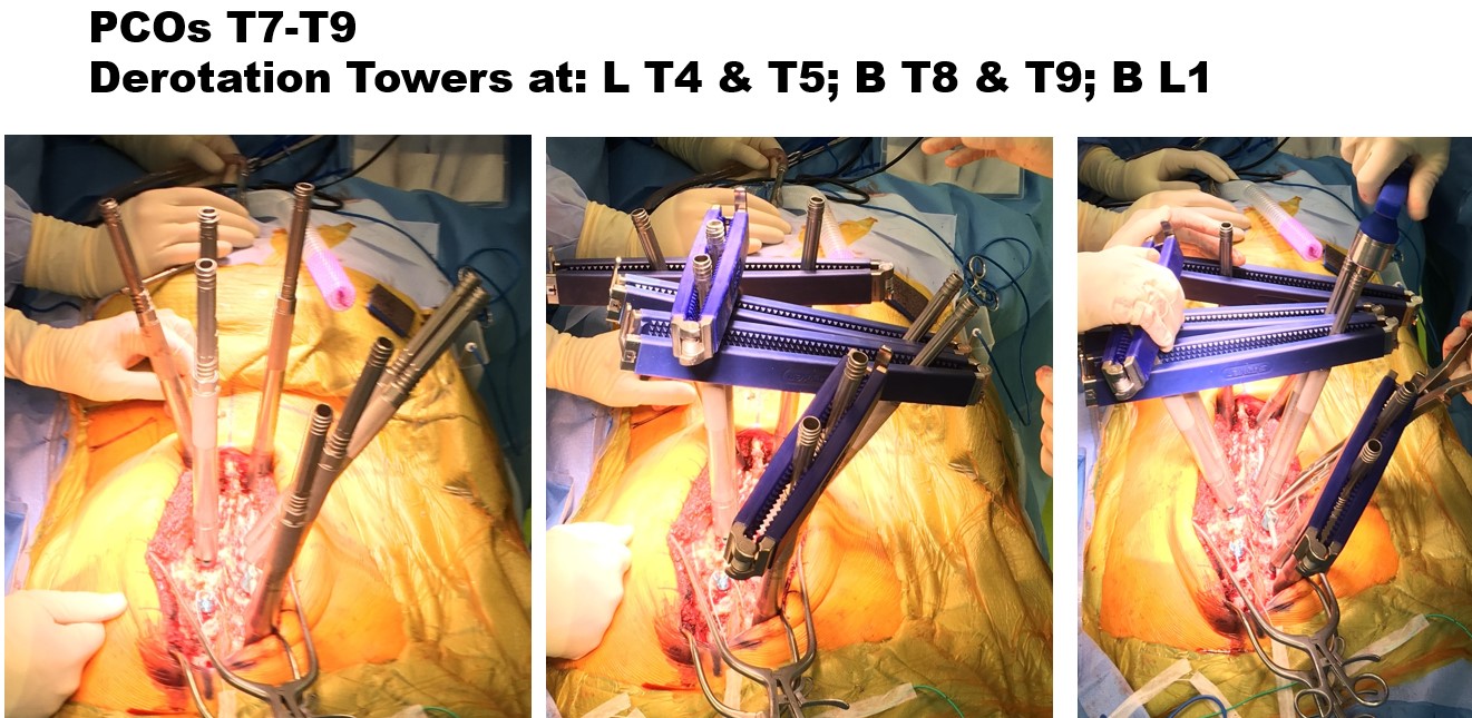
The silver towers or rods are attached to the screws (picture on left), and then these towers are connected together to improve strength of the spine fixation (middle picture). This connection process, for this patient, make 3 groups, one for each curve. These 3 groups of towers/screws are then rotated back toward normal (picture on right). You can see the different position of the middle group, relative to the other two groups, between the middle and right-sided pictures. Once rotated to the improved position (right picture) the screws are then tightened down, and much more work is done in surgery to 3-dimensionally improve and balance the spine.

These postoperative radiographs are one year after surgery. The patient is nicely balanced 3-dimensionally and has nicely improved.