What is a Vertebral Column Resection (VCR)?
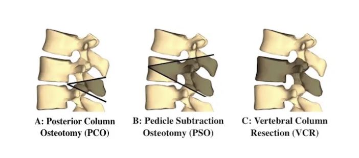
A VCR is complete surgical removal of one, or more,
vertebra. It is always combined with
posterior spinal fusion and instrumentation.
This means screws, rods, and possibly cages are used to hold the spine
in the new alignment while the spine undergoes bony fusion. Example C below.
A VCR is also called a Three-Column Osteotomy or a
Corpectomy.
When is a VCR
needed?
A VCR may be needed when a severe, rigid spinal deformity is
to be corrected. It can be used to treat hyperkyphosis (abnormal forward
curvature of the spine) or scoliosis (abnormal side curvature of the spine).
Why would more
than 1 vertebra be removed?
Removal of a single vertebra can permit 80+ degrees of
correction. Despite this fact a 2nd
vertebra may also need to be removed to achieve the desired correction safely.
What happens to
the gap between the vertebra after a VCR?
After a VCR the two ends of the spine can be moved in space
to correct the spine deformity. Ideally
there is no gap, and the vertebra can be moved so that there is bone-on-bone
contact. However, at times there is a
gap after the spine deformity is corrected.
The presence of a gap or hole is dependent upon the type and severity of
the spinal deformity. If there is a gap a “cage” is placed which struts from
the upper vertebra to the lower vertebra.
Why is a “cage”
sometimes placed?
A “cage” strengthens the surgery, by providing a strut from
vertebra to vertebra on the front side of the spinal cord. The usual spinal instrumentation is placed in
the back of the spinal cord. So a “cage”
gives 2 areas of stabilization for the spine.
This can increase the likelihood the instrumentation holding the spine
and a successful fusion of the surgery.
Below is a 10 year old female with Neurofibromatosis Type 1
and a progressive kyphosis. On the left
x-ray the vertebra with the red arrow pointing to it was removed. On the right, the blue arrow points to the
“cage” which was placed at the defect site, to support the spinal column in
front of the spinal cord.


Next week we will go over how we do this surgery.