the initial steps which are done, just before a VCR is performed. This post will talk about how a VCR is actually completed.
How is a Vertebral
Column Resection Performed?
After the incision, spinal exposure and placement of pedicle
screws the next important step is to place a rod across the VCR site (see below
at green arrow). This is important as a
VCR significantly destabilizes the spine, and not having 1 or 2 rods across the
VCR the spine can move, or subluxate, which can cause the spinal cord to not
function normal.

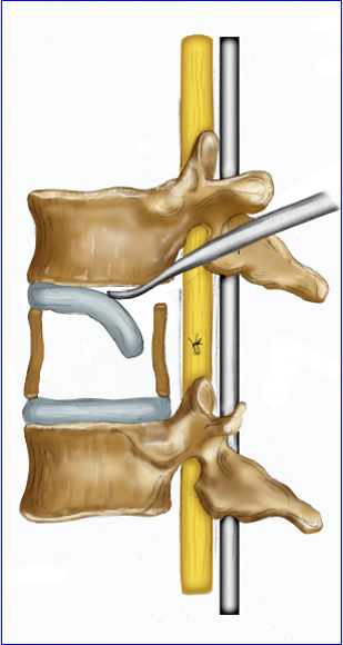
Next, the VCR step is to carefully remove the vertebra of
interest, piece by piece, working from the back of the vertebra to front. The
back, or roof, of the spinal column if first take off, to expose the spinal
cord (see blue arrow in the surgical photo and the red arrow in the drawing).


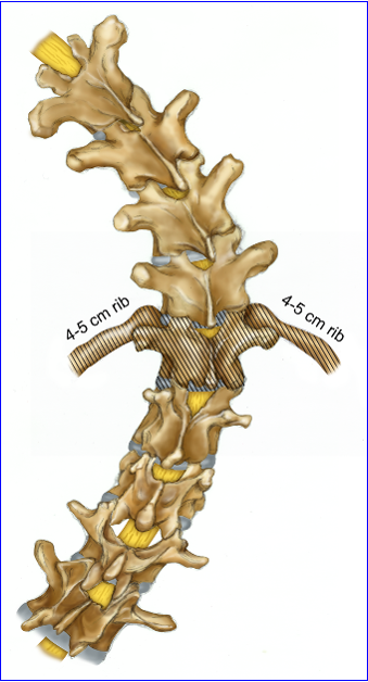
The 2 ribs connected to the vertebra of interest are identified
and exposed the medial 4-5 cm of the ribs are removed.
After the lamina is resected, retractors are placed around
the vertebral body to safely expose the bone and protect the vital structures
on the sides and in front of the spine (see below, red and black lines).

See the operative picture below for the retractor (green
arrow)

Next the pedicles (see red arrows in below diagram), the
column of bone which connects the back or roof of the spinal column to the
vertebral body are resected. This would leave only the vertebral body (#1).

Now the spinal cord can be seen on the back, left and right
side. The bone of the vertebral body is
the carefully removed (below) with curettes and drills.


After removal of the body…the discs on each side of the VCR
are then removed (see below).

Once the vertebral body is completely removed, the spinal
cord is a tube which bridges from one vertebral to the adjacent vertebra (green
arrow below).

It is now time to take the deformed spine and realign it to
a better position, which is done by bending the rod and compressing the spine
above and below the VCR defect site together (see below)
.

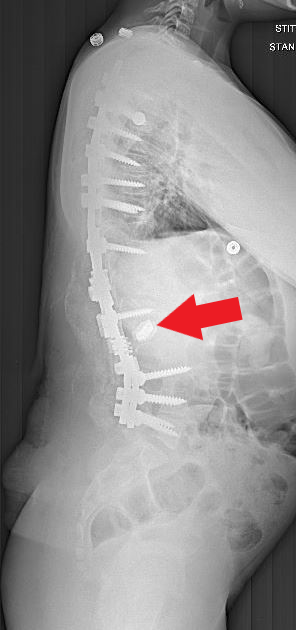
Sometimes the space can be completely closed down, bone on
bone. Other times a small “cage” is
needed to bridge the gap. This is
important from a spinal stability and healing of the bone fusion. A gap in the front can allow too much
movement of the spine and prevent the fusion from healing, causing the rods to
break or screws to move or pull out of the bone.

A cage was used in the surgical case shown below (red
arrows)

Once good spinal alignment is achieved, the spinal column is
stabilized with 2 or more rods, in its new and improved position.
All the implants (pedicle screws, hooks, cages and rods) help
to attain the new spine alignment, but also maintain it until the spine fusions
set up and is durable. The development
of a spine fusion can take several years to get hard and durable.