What is bone age?
Bone age is a method to determine the skeletal maturation
level of a growing person. The younger
the bone age, the more growth is ahead of them.
Why not just use
someone’s chronologic age?
The chronologic age of a growing individual, which is
calculated from their birth date to now, does not accurately estimate the
maturation of their bones. We all know
people who are “early bloomers” and “late bloomers”, those young men who
started shaving in 7th grade vs. those who kept growing until they
started college. There is wide variation
in bony maturation and growth when we look at someone’s chronologic age.
Why does bone age
matter?
We can get much more precise when we look at the actual
bones, and their growth plates, to get a better idea of someone’s future
growth. Remember, the more growth a
person with scoliosis has remaining the greater the likelihood the scoliosis
will worsen, and faster growth (during the pubertal growth phase) usually means
faster progression (worsening). So
better knowing how much growth is in the future helps plan treatment more
precisely.
How do you tell how
old are someone’s bones?
Radiographs/x-rays are used.
Many different parts of the body have been used for determination of
bone age, from the calcaneus (heel bone) to the olecranon (elbow) to the
humerus (shoulder) and the hand. Classically, spine surgeons have use the
Risser sign, which grades the iliac crest (hips) growth plates. Since this part of the body is seen on spine
radiographs/x-rays it doesn’t require any additional imaging. See below image,
the iliac crests are at the blue arrows.

Where does someone
look for the Risser sign?
The iliac bones (above, blue arrows), and there are two of
them, one on the left and one on the right side of the pelvis. The sit on each side of the sacrum and look
like Mickey Mouse ears.

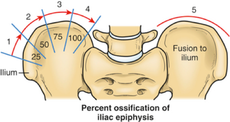
How is the Risser sign determined?
The Risser sign is from 0 to 5 (see below). The iliac apophysis (a growth center for the
iliac crest) is a thin strip of bone that forms on top of the iliac
crests. If there is no bone visualized
on the plain radiographs/x-rays then it is called a Risser 0. The strip of bone keeps growing back to cover
the entire iliac crest (Risser 4), and then fuses to the iliac crest (Risser
5). Once the Risser 5 stage is reached
the patient has completed skeletal growth.
Risser Sign
Stage
Description
0
Bony iliac apophysis not yet visible
1
Initial (<25%) ossification of the iliac
apophysis
2
From 25% to 50% ossification of the iliac
apophysis
3
From 50% to 75% ossification of the iliac
apophysis
4
More than 75% ossification of the iliac
apophysis
5
Iliac apophysis fuses to the iliac crest

Below is an example of a Risser 1-2 sign. See the very small amount of bone on the
outside part of the iliac crest (on the right side just below the “300” on the
radiograph/x-ray).

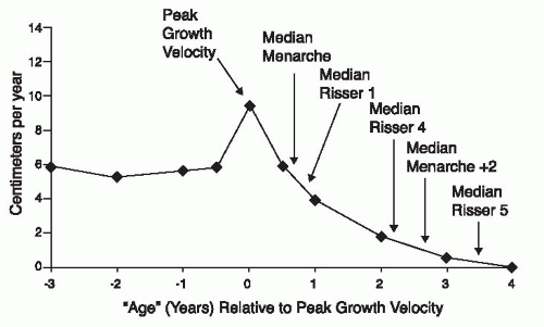
The below graph demonstrates the velocity of growth, with
year “zero” being the time of peak growth velocity. As you look to the right, after menarche
occurs, then Risser sign 1 occurs, almost a full year after peak growth
velocity. This is a weakness of the Risser sign, it doesn’t identify the peak
growth velocity, as it only occurs after it has happened.
To deal with this the Sanders grade was developed by Dr. Jim
Sanders, the Chair of Orthopaedics at the University of North Carolina.
Next blog post a better way to estimate skeletal
age will be presented: The Sander grading system.