To summarize the previous post, the pars interarticularis likely never developed normally for individuals who get a spondylolysis or pars fracture. With continued stress on this area during normal and athletic activities the abnormally-developed pars cannot handle the repetitive stresses applied and a crack develops.
If the pars is bone, why doesn’t it just heal like other fractures in the body? As demonstrated in the previous post the area of the pars is rather narrow, and the bone at the pars is mainly comprised of cortical bone (slow healing ability) rather than cancellous bone (fast healing ability). So, unlike other bones of the body which have a lot of cancellous or spongy bone, which heals faster, the pars has more cortical or dense bone, which heals slower. Normally our bone gets stronger as we apply stress to it, during our physical activities, as long as it is in a gradual fashion and the bone has time for it to recover from the stress and make new, stronger bone. However, if the bone is repetitively over-stressed, faster than it can heal, a crack or fracture can occur. We also see this in other areas of the body, such as the tibia or the foot in runners, and we call them “stress fractures”.

What symptoms are associated with a pars fracture/spondylolysis? Like a fracture in other areas of the body the main symptom is localized pain at the fracture site. Pain is typically worse with more strenuous activities, such as sports, and improves with rest. Other symptoms can also be present, such as pain which radiates to the buttocks and back of the thighs, which also gets worse with activities. Paralysis/weakness of the lower extremities and bowel/bladder dysfunction do not occur from an isolated spondylolysis.

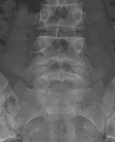
What is the first step in diagnosing a pars fracture/spondylolysis? The first step is a simple two view radiographic series in the AP and lateral projections. Additional oblique views can be helpful.
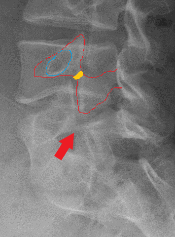
The below radiographs are of a 14 year old female with 18 months of low back pain

It is not easy to see but at the tip of the red arrow there is decreased bone density, which is indicative of a pars fracture/spondylolysis, but is not definitive.
Right ObliqueLeft Oblique


The above two oblique radiographs demonstrate bilateral (both sides) pars fractures or spondylolysis.
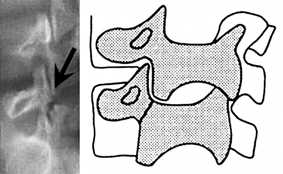
The below oblique demonstrates the “Scotty Dog” of the posterior elements of the spine. The pars fracture or spondylolysis looks like the dog is wearing a collar.
Can a pars fracture/spondylolysis always been seen on plain radiographs? The simple answer is: no. If the symptoms are for less than 2 months the plain radiographs may be negative. Sometimes more advanced imaging is necessary, such as CT scans, MR imaging or bone scans.
Below is a CT scan demonstrating bilateral pars fractures/spondylolysis (black arrows) at L5
Axial image Left pars Right pars


Next post will discuss treatment options for pars fractures/spondylolysis.