Surgical Case
The case presented is a 13 year old male with NF-1 who has a severe, progressive,
painful kyphoscoliosis.
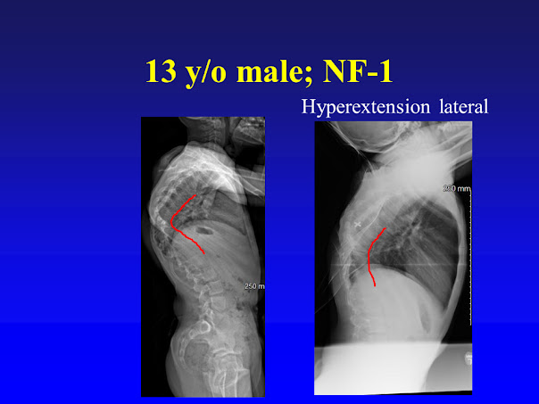
There is
some inherent spinal flexibility as the thoracic kyphosis of 91 degrees
improves when he lays on his back and hyperextends.
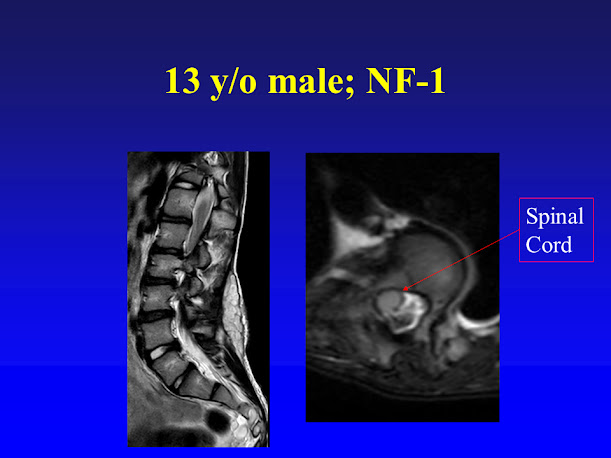
The below
selected MRI cuts demonstrates he does not have significant dural ectasias
which could complicated surgery. The
axial MRI cut shows the spinal cord very eccentric in the canal, resting
against the pedicle. The spinal cord is
slightly out-of-round, which elevates the risk of neurologic issues during
surgery.
The below coronal
CT scan cuts nicely shows the apex of the scoliosis having very abnormal
vertebra. Instead of being rectangular
they are trapezoidal or triangular, which makes the scoliosis have a very tight
turn.

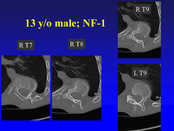
The below
axial CT scan cuts demonstrates the very abnormal pedicles. Several of these pedicles (R T7, R T8 and R
T9) are very difficult to place straight pedicle screws. The reason these can be cannulated safely is
due to the bone being malleable or bendable, and the pedicles can be bent
straight (within reason).
The patients
underwent 4 weeks of in-patient halo-gravity traction, with a maximum traction
weight of 28 lbs. Despite the spine improving above and below the apex of the
scoliosis, and the kyphosis improving, there still was a stiff apex.

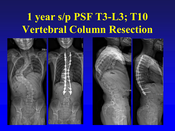
Surgical treatment
was a T3-L3 posterior spinal fusion and a T10 vertebral column resection, which
means the entire T10 vertebra was removed.
This technique disconnects the spine and dramatically increases the
flexibility of the spinal deformity.
After the spine was straightened a metal cage was placed in the front to
help attain and maintain correction.
The patient
is one year out from surgery and his doing well without pain.